[ad_1]

It is no secret that our lungs play a significant position in our every day lives—making certain we will breathe, fend off infections, and adapt to numerous challenges. Regardless of their significance, the organs nonetheless puzzle many medical consultants, particularly after they’re affected by ailments. Whereas conventional instruments like MRI and CT scans are useful when a affected person is experiencing a lung-related sickness, they will nonetheless fall brief in offering the detailed, real-time info wanted to know the intricacies of lung well being.
Enter the groundbreaking innovation generally known as the crystal ribcage. Developed by researchers in Boston College’s Faculty of Engineering, Pulmonary Middle, Middle for Multiscale and Translational Mechanobiology, and Neurophtonics Middle, the expertise is poised to revolutionize not solely our understanding of lung operate but additionally holds immense potential for different organs and coverings.
In new analysis, published this month in Nature Strategies, the crystal ribcage acts as a transparent, protecting protect for a mouse’s lungs, permitting scientists to get an in depth view of how these organs work in real-time, and at a mobile degree. What makes this expertise particular is that it would not disrupt the lung’s pure processes—respiratory and blood circulation proceed as regular whereas the researchers observe.
On this Q&A, senior creator Dr. Hadi Nia discusses the crystal ribcage, the way it’s reshaping our understanding of lung analysis, and its potential makes use of past the lungs.
What’s the major problem highlighted within the analysis in the case of understanding lung well being?
The lung is the location of many deadly pathologies similar to major and metastatic cancers, respiratory infections and each obstructive and restrictive ailments that affect its capabilities on the mobile degree. With current imaging modalities similar to MRI, CT, and histological analyses, dynamic single cell occasions on the early phases of illness development, such because the interactions of immune cells with most cancers cells and micro organism, can’t be resolved.
With the crystal ribcage, we will for the primary time picture the mouse lung all the way down to mobile ranges whereas it retains its physiological functions similar to respiration, circulation of blood, and immune exercise. We are actually in a position to research many lung ailments on the earliest steps of illness initiation.
How does the crystal ribcage differ from conventional strategies like MRI and CT scans when learning the lungs?
MRI and CT permit the visualization of the entire lung, however their spatial and temporal resolution are low, and single air sacs (generally known as alveolus), single capillaries, and mobile occasions similar to migration of immune and most cancers cells can’t be resolved. Crystal ribcage then again, permits using optical microscopy by changing the precise ribcage made out of bone and muscle and therefore blocking the sunshine, by a clear rib made kind biocompatible supplies.
Since crystal ribcage has the identical geometry and materials properties of the particular rib, the lung can keep its physiological capabilities as being imaged by an optical microscope. If mixed with a quick sufficient microscope, the dynamic occasions similar to mobile trafficking and blood stream could be imaged in real-time when the lung is in motion. This expertise principally opens the black field of the lung in well being and illness, and offers real-time views of the lung that had been by no means seen earlier than.
Are you able to describe how the crystal ribcage works to allow shut remark of a mouse’s lung?
The crystal ribcage design is knowledgeable from the microCT of the native mouse ribcage to make sure that the lung can operate physiologically contained in the crystal ribcage. The lung inside crystal ribcage is ventilated both by positive-pressure to simulate mechanical air flow, or with negative-pressure to simulate spontaneous respiratory. The lung can be circulated with media or blood to supply vitamins to the lung cells. The crystal ribcage is then positioned on the microscope stage and the lung is imaged on the decision of curiosity.
Along with the imaging capabilities that crystal ribcage offers, we’re in a position to intervene in some ways within the respiration or circulation, similar to including medicine, or altering the lung physiological parameters (e.g., simulating train by growing respiratory fee and depth), and research the following adjustments within the lung structure-function. This controllability when mixed with imaging capabilities make crystal ribcage a transformative expertise to review many key lung ailments.
Along with lung analysis, what different organs would possibly profit from the applying of the crystal ribcage expertise?
We now have used the crystal ribcage to picture the guts at the side of the lung at elevated vascular pressures. Such experiments will permit additional probing of circulation-respiration coupling in illness similar to pulmonary hypertension or arrhythmia sooner or later. With the success and enthusiasm that now we have generated, future instructions in Nia Lab embrace translating comparable concepts to the mind by fabricating a “crystal cranium” to visualise the entire mind in motion.
How would possibly the crystal ribcage expertise advance research associated to organ transplants and regenerative drugs?
Crystal ribcage advances these research in two methods:
- Offering a physiological atmosphere for the transplant or engineered tissue the place the pattern could be imaged on the mobile decision in real-time.
- Permitting intervention by altering the physiological parameters, administration of therapeutic brokers, and monitoring the following alteration within the lung.
As one instance, there’s lively analysis on how lengthy we will maintain the human lung viable in between the donor-receiver surgical procedures. This time, presently at 6–8 hours, if prolonged, could be extraordinarily invaluable because it permits storing and transporting the organ to different transplantation websites. Our crystal ribcage permits monitoring the ex vivo lung on the optical decision, and therefore permits the researchers to higher perceive why the human lung can’t be maintained exterior the physique for greater than 6–8 hours.
As one other instance, there’s an lively line of analysis on lung tissue engineering. Regardless of the progress on engineering the lung, there isn’t any dependable solution to consider the operate of the lung. Crystal ribcage permits this analysis on the cellular level whereas permitting the engineered lung operate in a physiological atmosphere.
How does the crystal ribcage contribute to a greater understanding of lung ailments, improvement, and getting old?
The crystal ribcage permits three-dimensional (3D) imaging over your complete floor of the lung, whereas uniquely combining the advantages of in vivo mouse fashions (mobile range, 3D lung structure, respiratory and circulation) and in vitro organ-on-chip fashions (imaging benefits and in depth, exact management over microphysiology). We principally can see how illness development and getting old have an effect on the lung structure-function at single cell decision and in real-time.
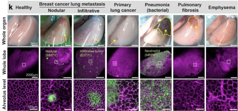
As examples, we utilized the crystal ribcage to probe transforming of alveolar and capillary capabilities in mouse fashions of major and metastatic lung most cancers, bacterial an infection, pulmonary fibrosis, emphysema, and acute lung harm. We recognized the earliest stage of tumorigenesis when alveolar construction–capabilities are compromised, probed the dynamic circulatory capabilities of capillaries transformed by tumor development and pneumonia and demonstrated a dramatic and reversible mechano-responsiveness in immune cell migration within the lung.
As one other instance, it’s well-known that getting old is main threat issue for pneumonia. Nevertheless, we have no idea precisely why. In collaboration with Joseph Mizgerd, the Listing of Pulmonary Middle, we are actually using crystal ribcage to higher perceive how getting old might have an effect on the pneumonia development or decision.
That is all however a tip of the iceberg and we’re excited to collaborate with the analysis neighborhood to additional our collective data of illness development, therapeutic results, and getting old of the lung because it capabilities.
Who’re your key analysis collaborators?
Our analysis collaborators span each the Charles River and Medical campuses and are a part of the BU Pulmonary Middle, BU Middle for Multiscale and Translational Mechanobiology, and BU Neurophtonics Middle. Key investigators who supported our analysis are Dr. Bela Suki, Dr. Joseph Mizgerd, Dr. Sarah Mazilli, Dr. Katrina Traber, and Dr. Giovanni Ligresti.
Extra info:
Rohin Banerji et al, Crystal ribcage: a platform for probing real-time lung operate at mobile decision, Nature Strategies (2023). DOI: 10.1038/s41592-023-02004-9
Quotation:
Q&A: ‘Crystal ribcage’ expertise pioneers new approaches to lung well being (2023, September 26)
retrieved 26 September 2023
from https://medicalxpress.com/information/2023-09-qa-crystal-ribcage-technology-approaches.html
This doc is topic to copyright. Other than any honest dealing for the aim of personal research or analysis, no
half could also be reproduced with out the written permission. The content material is supplied for info functions solely.
[ad_2]
Source link


Discussion about this post