[ad_1]

Credit score: Mayo Clinic Information Community
Ultrasound—a know-how that makes use of sound waves to supply a picture—is usually used to watch the event of a child because it grows inside its mom. However ultrasound imaging additionally can be utilized to research suspicious lots of tissue and nodules that could be cancerous.
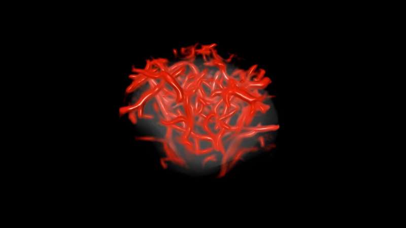
Tumors consist not solely of cancer cells but in addition a matrix of small blood vessels, or microvessels, that can’t be seen within the photographs produced by standard ultrasound machines. To resolve this drawback, physician-scientist Azra Alizad, M.D., and biomedical engineering scientist Mostafa Fatemi, Ph.D., teamed up at Mayo Clinic to design and examine a software that will enhance the decision of ultrasound imaging.
As demonstrated in research findings printed final April in European Radiologythey developed high-resolution ultrasound imaging software program, appropriate with many ultrasound machines, that might exponentially enhance each the element and high quality of photographs.
The investigational software program, which they’ve named quantitative high-definition microvessel imaging (q-HDMI), has been evaluated to seize high-resolution 2D and 3D photographs of microvessels as small as 150 microns, roughly twice the width of a human hair.
“If we will visualize and seize the microvessel within the earliest phases of cancerwe will higher diagnose and deal with it earlier, which improves the end result for the affected person,” says Dr. Alizad, who focuses on ultrasound know-how for most cancers imaging.
Synthetic intelligence helps detect what we can not see
As well as, the researchers recognized a sequence of biomarkers representing particular traits of tiny vessels, similar to form, sample, irregularity and complexity, and packaged them into an algorithm that may type the image data into benign or malignant lots.
“This know-how supplies a quantitative worth that reveals the chance of malignancy,” Dr. Fatemi says. “It is a software to extract info in a means that may be helpful to clinicians.”
Making use of it in follow
In a clinical study printed final November in Breast Most cancers Analysisthe researchers confirmed that their new q-HDMI software mixed with artificial intelligence (AI) was capable of detect a really small, 3-millimeter-wide, malignant breast most cancers mass comprised of miniscule vessels in a 40-year-old lady. Discovering and treating most cancers lesions when they’re this measurement earlier than they unfold, or metastasize, will be lifesaving.
“The questions in radiology actually are: benign or malignant? And if it is suspicious, how involved ought to we be?” Dr. Fatemi says.
To assist radiologists reply these questions, the researchers utilized their software program and algorithm to additional analyze suspicious breast lots from 521 sufferers who already had obtained standard ultrasound imaging. The outcomes have been astounding. The brand new know-how yielded a virtually 100% price of accuracy in figuring out malignant versus benign lots, whatever the tumor measurement.
Extra just lately, the researchers analyzed thyroid nodules from 92 sufferers. Thyroid nodules are frequent and it’s typically laborious to distinguish between cancerous and non-cancerous ones by means of imaging. But the prevalence of thyroid most cancers has elevated over the previous few a long time.
“With standard ultrasound photographs, physicians can diagnose whether or not a thyroid nodule is benign or malignant with solely about 35%–75% accuracy,” Dr. Alizad says. That is why physicians typically choose to carry out thyroid biopsies, which allow them to extra definitively decide if a thyroid nodule is a trigger for concern.
The researchers recognized 12 biomarkers that may differentiate benign from malignant thyroid tissues. They programmed the AI-powered algorithm with these biomarkers, which sorted the photographs and had an 84% price of accuracy. These outcomes have been printed within the journal Cancers and highlighted by the Nationwide Institutes of Well being.
“Whether it is most cancers, we positively need to know that. But when we will decide if a thyroid nodule is benign with out even having to do a biopsy, that’s even higher because it spares the affected person from the monetary and bodily burdens related to an pointless benign biopsy,” Dr. Alizad says.
The researchers see that their quantitative software might be significantly helpful in components of the world the place there’s restricted experience and assets, similar to rural areas and growing international locations.
Additionally they are collaborating with oncologists to allow them to make use of the q-HDMI software to raised monitor the effectiveness of most cancers therapies and assist them regulate therapies for particular person sufferers in actual time.
Extra info:
Melisa Kurti et al, Quantitative Biomarkers Derived from a Novel Distinction-Free Ultrasound Excessive-Definition Microvessel Imaging for Distinguishing Thyroid Nodules, Cancers (2023). DOI: 10.3390/cancers15061888
Quotation:
Advancing ultrasound microvessel imaging and AI to enhance most cancers detection (2023, October 21)
retrieved 21 October 2023
from https://medicalxpress.com/information/2023-10-advancing-ultrasound-microvessel-imaging-ai.html
This doc is topic to copyright. Other than any honest dealing for the aim of personal examine or analysis, no
half could also be reproduced with out the written permission. The content material is offered for info functions solely.
[ad_2]
Source link


Discussion about this post