[ad_1]

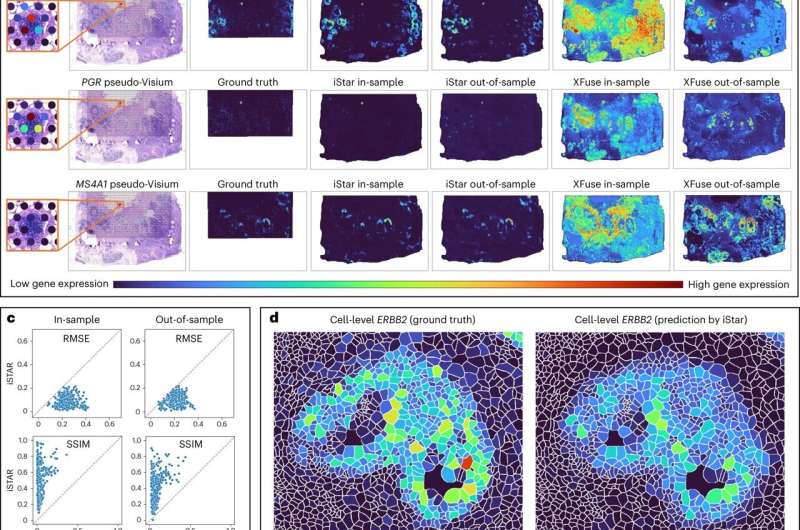
Workflow and super-resolution gene expression prediction accuracy of iStar. Credit score: Nature Biotechnology (2024). DOI: 10.1038/s41587-023-02019-9
A brand new synthetic intelligence software that interprets medical photos with unprecedented readability does so in a means that might enable time-strapped clinicians to dedicate their consideration to vital points of illness analysis and picture interpretation.
The software, known as iStar (Inferring Tremendous-Decision Tissue Structure), was developed by researchers on the Perelman College of Drugs on the College of Pennsylvania, who consider they will help clinicians diagnose and higher deal with cancers that may in any other case go undetected.
The imaging technique gives each extremely detailed views of particular person cells and a broader have a look at the total spectrum of how individuals’s genes function, which might enable docs and researchers to see cancer cells that may in any other case have been just about invisible. This software can be utilized to find out whether or not secure margins had been achieved via most cancers surgical procedures and routinely present annotation for microscopic photos, paving the best way for molecular illness analysis at that stage.
A paper on the tactic, led by Daiwei “David” Zhang, Ph.D., a analysis affiliate, and Mingyao Li, Ph.D., a professor of Biostatistics and Digital Pathology, was printed immediately in Nature Biotechnology.
Li stated that iStar has the power to routinely detect vital anti-tumor immune formations known as “tertiary lymphoid buildings,” whose presence correlates with a affected person’s seemingly survival and favorable response to immunotherapy, which is commonly given for most cancers and requires excessive precision in affected person choice. This implies, Li stated, that iStar may very well be a strong software for figuring out which sufferers would profit most from immunotherapy.
The event of iStar was taken on as a part of the sphere of spatial transcriptomics, a comparatively new area used to map gene actions throughout the house of tissues. Li and her colleagues tailored a machine studying software known as the Hierarchical Imaginative and prescient Transformer and skilled it on customary tissue photos.
It begins by breaking down photos into completely different levels, beginning small and searching for high-quality particulars, then shifting up and “greedy broader tissue patterns,” based on Li. A community guided by the AI system inside iStar makes use of the knowledge from the Hierarchical Imaginative and prescient Transformer to then take in all of that data and apply it to foretell gene actions, usually at near-single-cell decision.
“The facility of iStar stems from its superior methods, which mirror, in reverse, how a pathologist would research a tissue pattern,” Li defined. “Simply as a pathologist identifies broader areas after which zooms in on detailed mobile buildings, iStar can seize the overarching tissue buildings and likewise deal with the trivialities in a tissue picture.”
To check the efficacy of the software, Li and her colleagues evaluated iStar on many several types of most cancers tissue, together with breast, prostate, kidney, and colorectal cancers, combined with wholesome tissues. Inside these exams, iStar was capable of routinely detect tumor and cancer cells that had been laborious to establish simply by eye. Clinicians sooner or later could possibly choose up and diagnose extra hard-to-see or hard-to-identify cancers with iStar appearing as a layer of help.
Along with the scientific potentialities introduced by the iStar method, the software strikes extraordinarily shortly in comparison with different, related AI instruments. For instance, when arrange with the breast most cancers dataset the group used, iStar completed its evaluation in simply 9 minutes. In contrast, one of the best competitor AI software took greater than 32 hours to provide you with an analogous evaluation.
Which means iStar was 213 instances sooner.
“The implication is that iStar will be utilized to numerous samples, which is vital in large-scale biomedical research,” Li stated. “Its pace can be vital for its present extensions in 3D and biobank pattern prediction. Within the 3D context, a tissue block could contain lots of to hundreds of serially minimize tissue slices. The pace of iStar makes it potential to reconstruct this enormous quantity of spatial information inside a brief time frame.”
And the identical goes for biobanks, which retailer hundreds, if not hundreds of thousands, of samples. That is the place Li and her colleagues are subsequent aiming their analysis and extension of iStar. They hope to assist researchers acquire higher understandings of the microenvironments inside tissues, which might present extra information for diagnostic and therapy functions shifting ahead.
Extra data:
Daiwei Zhang et al, Inferring super-resolution tissue structure by integrating spatial transcriptomics with histology, Nature Biotechnology (2024). DOI: 10.1038/s41587-023-02019-9
Quotation:
New AI software brings precision pathology for most cancers and past into faster, sharper focus (2024, January 2)
retrieved 3 January 2024
from https://medicalxpress.com/information/2024-01-ai-tool-precision-pathology-cancer.html
This doc is topic to copyright. Aside from any truthful dealing for the aim of personal research or analysis, no
half could also be reproduced with out the written permission. The content material is offered for data functions solely.
[ad_2]
Source link


Discussion about this post